Premature degeneration of the intervertebral discs, like hemivertebrae, is an all-too-common problem in French Bulldogs. In Sweden, research indicates that Frenchies are second only to Dachshunds in the incidence of disc disease, proportionate to the breeds' population sizes. No data are available for the frequency of the condition in the United States Frenchie population, but it seems to be high here as well.
Researchers believe that the breeds most commonly affected are the "chondrodystrophic" ones in which an abnormal development of the bones results in various skeletal disproportions, such as short legs and noses. In these breeds, chondrodystrophy predisposes the discs to be prematurely converted to an abnormal type of tissue, whose consistency is unsuitable for the discs' function. Then, in the parts of the back most subject to mechanical stress, these degenerated discs are liable to rupture or protrude.
It is likely that a tendency toward disc disease, like hemivertebrae, is a side effect of the developmental defects that we select for as characterizing our lovely breed. But some lines of Frenchies probably have a higher-than-average incidence of the ailment superimposed on this built-in breed tendency. Inbreeding and line-breeding would increase the probability of an undesirable characteristic just as they would increase the incidence of the desirable ones.
As with other inherent breed problems that accompany the desirable traits, a perfect conformation may be too expensive if its cost is the health or the life of the Frenchie. It might be wise to reconsider the breed standard periodically to see whether a minor change in it could produce a healthier breed.
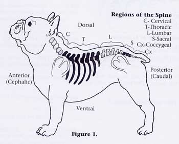
A first step toward coming to terms with this or any health problem is to educate Frenchie Friends breeders and pet owners alike as to the nature of the disease, its clinical signs, prognoses, and treatment. Underlying those beautiful folds and wrinkles of the Frenchie back is the vertebral column, consisting of 7 cervicals, 13 thoracics, 7 lumbar, 3 sacral, and a variable number of coccygeal (tail) vertebrae. The latter group is greatly reduced and generally abnormally formed in our little screwtailed friends (FIG. 1).
Between the ventral solid parts (bodies) of each pair of adjacent vertebrae is a flexible, cushion-like disc composed of a tough, outer fibrous envelope (the annulus) containing a soft, jelly-like inner mass (the nucleus). Either with aging or due to premature disc degeneration, dehydration of the disc may occur, resulting in a loss of elasticity.
The nucleus of the disc may then thicken, become fibrous, and finally calcify. These degenerative changes, generally present to some extent in old dogs of all breeds, often occur prematurely in the chondrodystrophic breeds.
Although the changes in the disc may be seen in x-rays as a narrowing of the intervertebral space, plus the presence of calcium deposits in the space, the degeneration of the disc does not itself generally cause clinical signs.
The problems arise when part or all of the disc is squeezed out from between the vertebrae and protrudes dorsally into the adjacent spinal canal, causing inflammation and/or compression of the spinal cord, and sometimes accompanied by bleeding from blood vessels in the tissues surrounding the spinal cord.
There may be a bulging out of the annulus, extrusion of the nucleus of the disc from its annulus (like squeezing the pulp of a grape out of its skin), or even total displacement of the entire disc.
The terms disc herniation, protrusion, rupture, extrusion, and prolapse have all been applied to various types of disc problems. Because these terms are not consistently used, we will refer to ANY movement of all or part of a disc out of its intervertebral space as a disc protrusion.
A degenerated disc may protrude slowly over some time, or rapidly; and it may happen for no apparent reason, or as a result of a trauma. Something as simple as a jump off a chair can dislodge or protrude a diseased disc, not that Frenchies are ever allowed on furniture, of course.
Protrusion usually occurs dorsally in the midline, into the spinal canal containing the spinal cord (FIG. 4). A ventral protrusion is rare because the annulus is thicker ventrally and is also reinforced ventrally by a strong ligament. The specific discs most often affected are those thoracolumbar discs between vertebrae T11 to L2 (where it is estimated that 60 percent of all disc protrusions occur), and in the cervical area, especially in the discs between vertebrae C2 to C4 (FIG. 2). The nine discs located between vertebrae T1 - T10 rarely protrude because they are reinforced dorsally by a ligament associated with the ribs which are attached to these vertebrae.
When disc degeneration is a consequence of age (in dogs ten years old or older), the cervical discs are the ones most often affected. In the premature condition, as often seen in Frenchies, both the cervical and the thoracolumbar discs show a high incidence of degeneration, with the latter being more often affected.
Protrusion of prematurely degenerated discs is most often seen in dogs that are three to five years old, though degenerative changes in the discs have been found to occur as early as four months of age. Both sexes are affected equally.
Clinical Signs of Intervertebral Disc Disease in French Bulldogs
The location of the protruding disc and the extent of the resulting spinal cord injury determine what signs the dog will show. The canal containing the spinal cord is much larger in the cervical region so that there is a relatively large space around the cord.
Thus, the degree of cervical cord compression is generally less severe and produces less serious signs than in the thoracolumbar area, where the spinal cord occupies about 80 percent of the available space in the spinal canal.
Protrusion of a cervical disc generally produces pain, but little if any muscle weakness or paralysis. The dog will walk with its head extended straight forward, and the neck muscles stiff and contracted. If the neck is manipulated, the pain is severe; and during an acute attack, the dog's body is rotated. If muscular involvement occurs, it will generally cause "knuckling" or stumbling of the forelimbs, especially when the involved disc is one of the more posterior ones in the cervical group.
In the thoracic and lumbar regions, the smaller diameter of the canal is likely to result in more serious cord compression, with a much greater likelihood of hind limb paralysis. In addition to directly compressing the spinal cord, a protruding disc may also cause inflammation and swelling of the cord, and/or hemorrhage by damage to the blood vessels in the tissues surrounding the cord.
Various sensory and motor "circuits" are contained in specific parts of the spinal cord and the type of paralysis, plus the presence or absence of pain, are clues from which the extent of damage to the cord can be deduced. Figure 3 illustrates a cross-section through the spinal column, canal, and cord. Numbers 1, 2, and 3 three demonstrate the locations of three important circuits within the cord. Numbers 1 and 2 represent motor circuits (i.e. controlling muscle movement), while number 3 represents the pain circuit. If a protruding disc causes a cord lesion or damage that extends into the cord as far as line B, interrupting circuits numbers 1 and 2, the hind limbs will lack muscle tone and will show flaccid paralysis.
Pain will still be present. In the most serious case, the cord damage extends all the way to line C, and affects all three circuits; in this case, the hind limbs will show a flaccid paralysis, but the loss of the pain circuit in an area no. 3 will result in an absence of pain.
This combination of signs indicates the most severe cord damage, with the least favorable outlook for recovery.
Intervertebral Disc Disease in French Bulldogs Treatment
The more rapidly the condition is diagnosed and treated, the better the chance for recovery, especially when the signs indicate that the cord injury is of the less extended type.
Drug therapy with corticosteroids and other anti-inflammatory agents may help reduce the inflammation and swelling of the cord; when coupled with pain-relieving drugs, improvement may be seen immediately.
However, relief of pain may reduce the dog's cautiousness and allow it to move around enough to further protrude the disc. It is a good idea to cage the dog to restrict movement during its convalescence. Most Frenchies love to rest anyway, so this doesn't really work a hardship on them. Physical therapy to help maintain muscle tone may include massage and manipulation of the limbs, whirlpool baths, and warm pads applied to the back.
Ultrasound may also help by increasing blood flow to the affected area and often relieves the muscle spasms that cause much of the pain.
Good nursing care should also include keeping the skin clean and dry and can prevent or minimize pressure ulcers of the skin (bed sores). If there is a loss of bladder and bowel control, the bladder should be emptied twice daily by manual pressure to help prevent bladder infections. Enemas may be given to prevent fecal retention. Any or all of these measures may be necessary to support the dog during its recovery if the condition is treatable.
However, if the condition does not respond to either medical or surgical treatment, euthanasia would be the kindest option. Pain and paralysis are no life for any dog ... especially a Frenchie. If surgery is indicated (on which point the owner should consider the opinion of his veterinarian, and may wish to consult a specialist for a second opinion), two surgical approaches are possible. One method for surgically relieving the pressure on the spinal cord is fenestration, in which the protruding disc material is removed from the intervertebral space using a needle or a scraper.
This may be done either by a ventral approach or by a lateral one (FIG. 4) and is more commonly done in cervical disc protrusions. An opening is made in the outer annulus of the disc, and its nucleus is removed through the opening. In this method, the spinal canal is not invaded, minimizing the danger of further injuring the cord.
Another surgical treatment, more often used to decompress the cord in the thoracolumbar disc protrusion, involves the removal of half of all of the dorsal arch of the vertebra (hemilaminectomy or laminectomy). Fig. 4 shows the portion of the vertebra removed. If paralysis is present, surgery may be justified if the animal's overall condition seems to warrant it. In the thoracolumbar area, where the canal is smaller and cannot accommodate as much swelling, decompression should be done within five to seven days, the sooner the better. It may be delayed longer in the cervical area.
Let us hope that by educating breeders and pet owners to recognize and deal with health problems in our breed, we may see a continual improvement in the soundness of our pups as well as in their conformation.
If breeders will continue to communicate openly with one another about potential problems in their lines as well as about desirable traits and outbreed occasionally to reduce the incidence of problems, the entire Frenchie breed will benefit in the long run. We must consider health and longevity to be as important a breed characteristic as bat ears and wrinkles; otherwise, we are not really our Frenchies' Friend.
From "Healthy Frenchies: an Owner´s Manual". Published by ArDesign. The USA.1998
First published in the French Bulletin, Vol. 3 No 2.1984


